
October 15, 2007
Should You Be Afraid Of Diagnostic X-Rays?
By Michael D. Shaw
Articles have been appearing in medical and dental trade magazines regarding patients who are refusing X-rays. In fact, the websites of many dentists devote a page or two to allaying patient fears, and noting how essential this diagnostic tool really is.
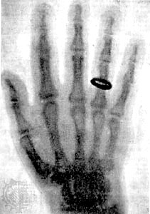
The discovery of X-rays dates back to 1895, when German physicist Wilhelm Konrad Röntgen was examining the effects of electron beams in electrical discharges through low-pressure gases. He soon observed that opaque objects placed between the radiation source and a screen, that would fluoresce in the presence of the rays, were effectively transparent. In 1896, he published the famous and dramatic image of the bones of a human hand.
Some time later it was determined that X-rays are electromagnetic radiation of extremely short wavelength (about 1000 times shorter than that of visible light) and high frequency, with wavelengths ranging from 10-12 to 10-8 meters and corresponding frequencies from 1016 to 1020 hertz (Hz).
X-rays (also called radiographs) are most popularly taken to:
- Determine whether a bone is chipped, dislocated or fractured
- Evaluate joint injuries and bone infections
- Diagnose and monitor the progression of degenerative conditions, such as arthritis and osteoporosis
- Detect scoliosis, and other spinal defects
- Locate dental problems such as cavities, abscessed teeth, and other tooth and jaw abnormalities
- Screen for lung and heart diseases
- Locate objects that may have been accidentally swallowed by a child
- Detection and diagnosis of cancer
While few would deny that X-ray and its more advanced cousin computed tomography (CT) are essential tools of modern medicine, a selective amnesia akin to those against vaccinations—after previously killer infectious diseases have been controlled—poisons the minds of a vocal few. Unfortunately, these few can draw support from a potpourri of radiation fears, cancer fears, pseudo-science, and the legitimate danger of large radiation exposures.
Indeed, you may have heard that even the smallest amount of radiation might cause cancer. Or, that the risk of causing a fatal cancer from a single chest X-ray is 10 times greater than the risk of dying in a commercial airline flight. Or, that a CT scan of the kidneys has a greater risk of inducing a fatal cancer than a cigarette smoker has of dying from any cancer.
Sadly, these nonsensical statements—and others like them—derive from the exposure model known as linear-nonthreshold dose-response. It assumes that there is no safe dose of radiation, and that the risk of getting cancer or genetic damage increases along with radiation exposure. Of course, the most immediate problem with this is that for most people, the greatest source of exposure is the natural environment, and regulatory levels are being set below the background.
The EPA advocates a standard for all radiation exposure from a single source or site at 15 millirem a year. A standard chest X-ray, in comparison, gives about 10 millirem to the chest, which is equivalent to 1 or 2 millirem to the whole body. The Nuclear Regulatory Commission sets its single source standard at 25 millirem a year. Yet, the natural level of background radiation in the United States, on average, is about 350 millirem a year, and in some areas of the country it is many times higher than that.
Listen to Dr. John Boice Jr., a radiation expert, who is the scientific director of the International Epidemiology Institute:
“In New York, for example, people absorb about 100 millirem of radiation each year from cosmic rays alone. In Denver, exposure from cosmic rays averages 200 millirem a year and natural variation in radiation exposure is many times the amounts of radiation that are being disputed by regulatory agencies. We eat, breathe and drink low levels of radiation.”
Another problem with linear-nonthreshold dose-response is that it was based on the rather simplistic 1927 conclusions of geneticist Dr. H. J. Muller, who elicited gene mutations in fruit flies by bombarding them with X-rays. The number of mutations in fruit flies was proportional to the dose of X-rays. However, Muller used extremely high levels of X-rays, and caused mutations, not cancer.
Moreover, while cancer etiology can be tied in with initially high and proportionately higher dosages of radiation, about one-third of the population gets cancer. The huge difficulty is finding any evidence that low doses of radiation cause cancers that would not have otherwise occurred. Bear in mind that even in the cohort of survivors of Hiroshima and Nagasaki, few excess cancers have been detected.
Certainly, there are special situations such as pregnancy, where one has to be careful, and excellent guidelines are given on the RadiologyInfo website, developed and funded by the American College of Radiology (ACR) and the Radiological Society of North America (RSNA).
Don’t let unreasonable fears, exacerbated by misguided Luddites, prevent you from obtaining the potentially lifesaving benefits of X-ray diagnostic technology.